Cardiovascular MRI analysis, free in your browser or on your desktop.
FreeCMR reviews, measures, and quantifies a multiparametric CMR study: cine function, tissue characterisation, flow, perfusion, and 3D. Run the Lite version instantly in your browser, or install the full version via the desktop app (coming soon).
Comparison
FreeCMR Lite vs. FreeCMR Desktop
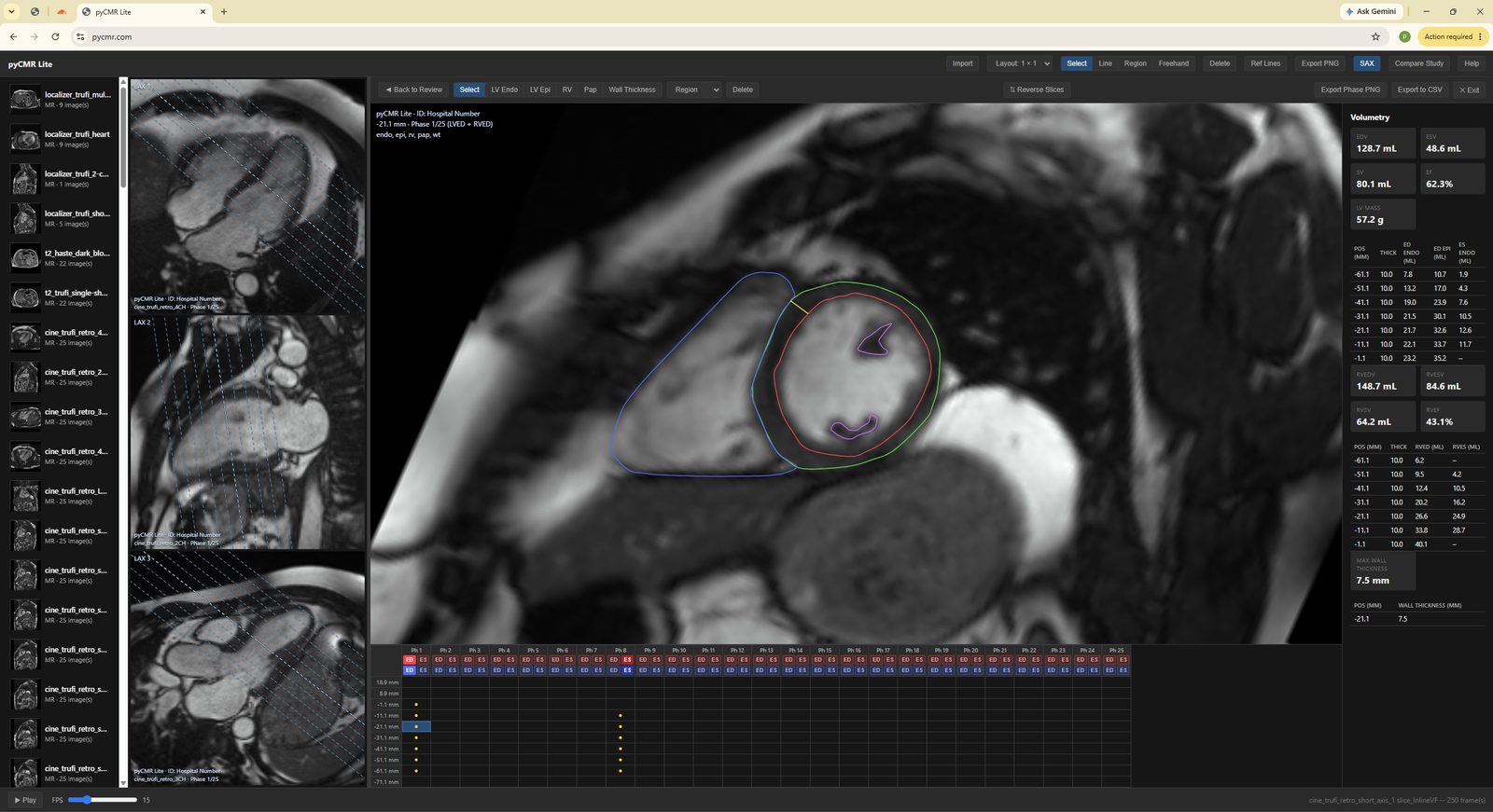
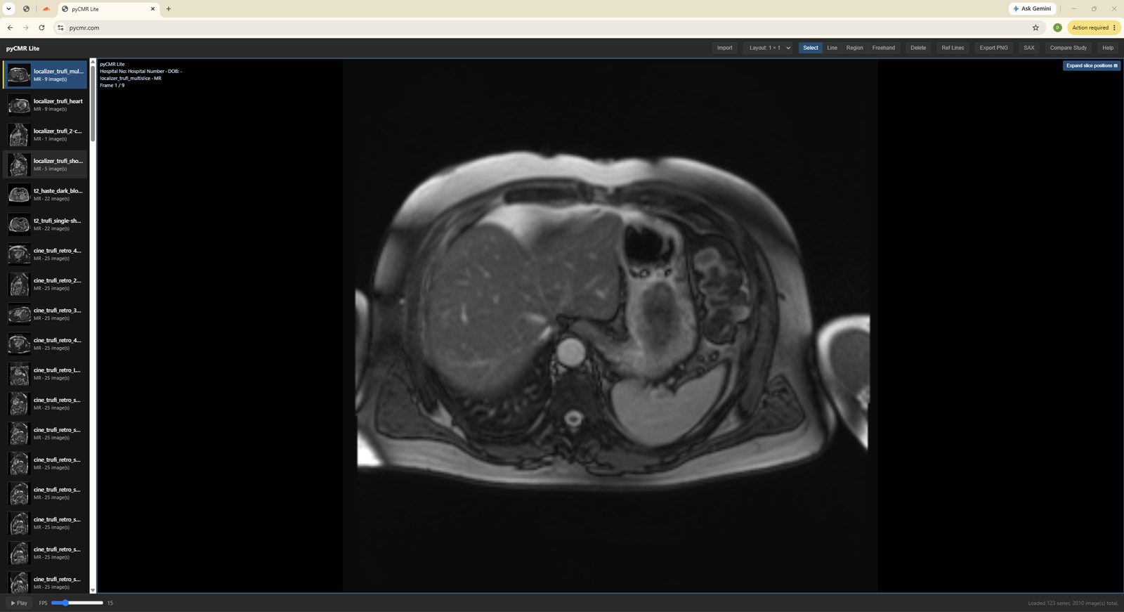
Import a folder of DICOM files: viewer, measurements, and a full short-axis workflow, right in your browser. Nothing you load is ever uploaded, it stays in that browser tab.
Open FreeCMR Lite →The full 10-module suite: viewer, short-axis, long-axis, T1/T2, T2*, perfusion, LGE scar quantification, flow, and 3D MPR. Installed locally, with a saved study database.
Get notified →FreeCMR Lite
A closer look at the browser app.
FreeCMR Desktop
Ten modules, one workspace.
every module ↓
Every module, in detail
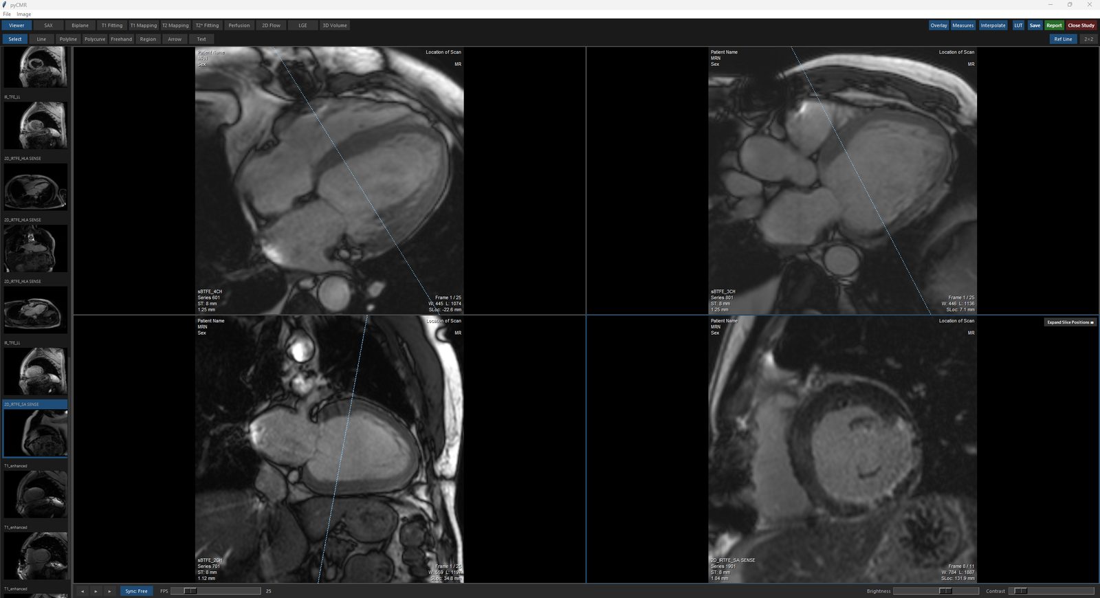
Viewer — main viewing module ▶
Cine playback, window/level, zoom/pan/rotate, general-purpose measurements, and study export and anonymisation.
- Multi-panel layout up to 4×2
- Line, Polyline, Polycurve, Region, Freehand, Arrow, and Text measurement tools
- Expand Slice Positions: review every slice of a stack at once, playing in lockstep, inside one panel
- Overlay, Measures, and LUT toggle on/off
- Anonymised DICOM/NIfTI export, plus CSV export of measurements
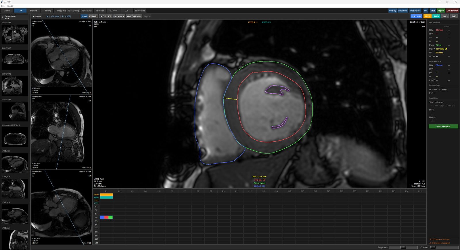
SAX — short-axis review and analysis ▶
Short-axis function analysis: LV and RV contouring, wall motion grading, volumes and mass.
- Base-to-apex stack assembly with long-axis reference views
- 16-segment RWMA bullseye, click to grade regional wall motion
- EDV, ESV, EF, mass, and wall thickness, per slice and indexed to BSA
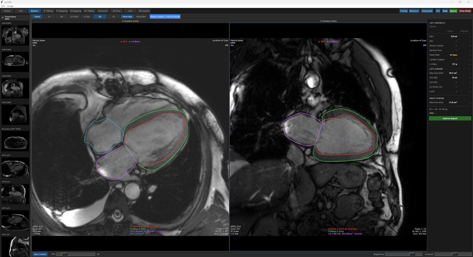
Biplane — LV, LA & RA from two long-axis views ▶
Left ventricular volumes by the biplane Simpson's method, plus left and right atrial area and volume, from a single screen.
- Simultaneous 4-chamber and 2-chamber contouring
- Linked phase marking across both views
- BSA-indexed results for LV, LA, and RA
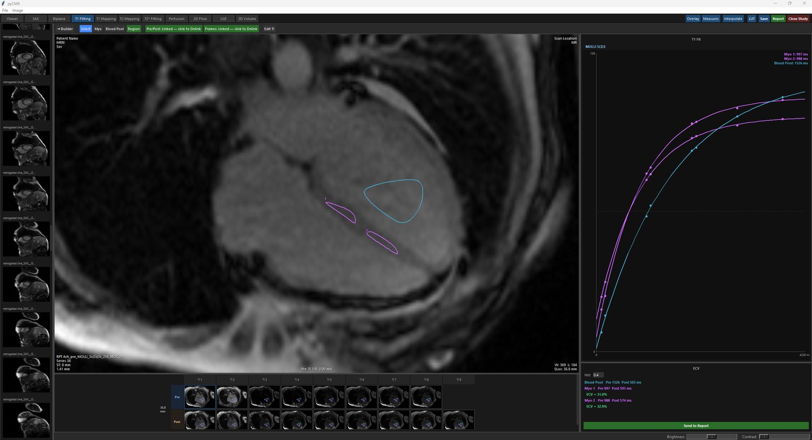
T1 Fitting — native & post-contrast T1, from source images ▶
Fits a T1 curve per ROI from raw multi-TI MOLLI/ShMOLLI source images, for native (Pre) and post-contrast (Post) acquisitions, with ECV.
- Automatic MOLLI/ShMOLLI sequence detection, overridable
- Pre/Post slice matching by position
- ECV from a haematocrit value and paired T1 fits
- Siemens and GE supported.
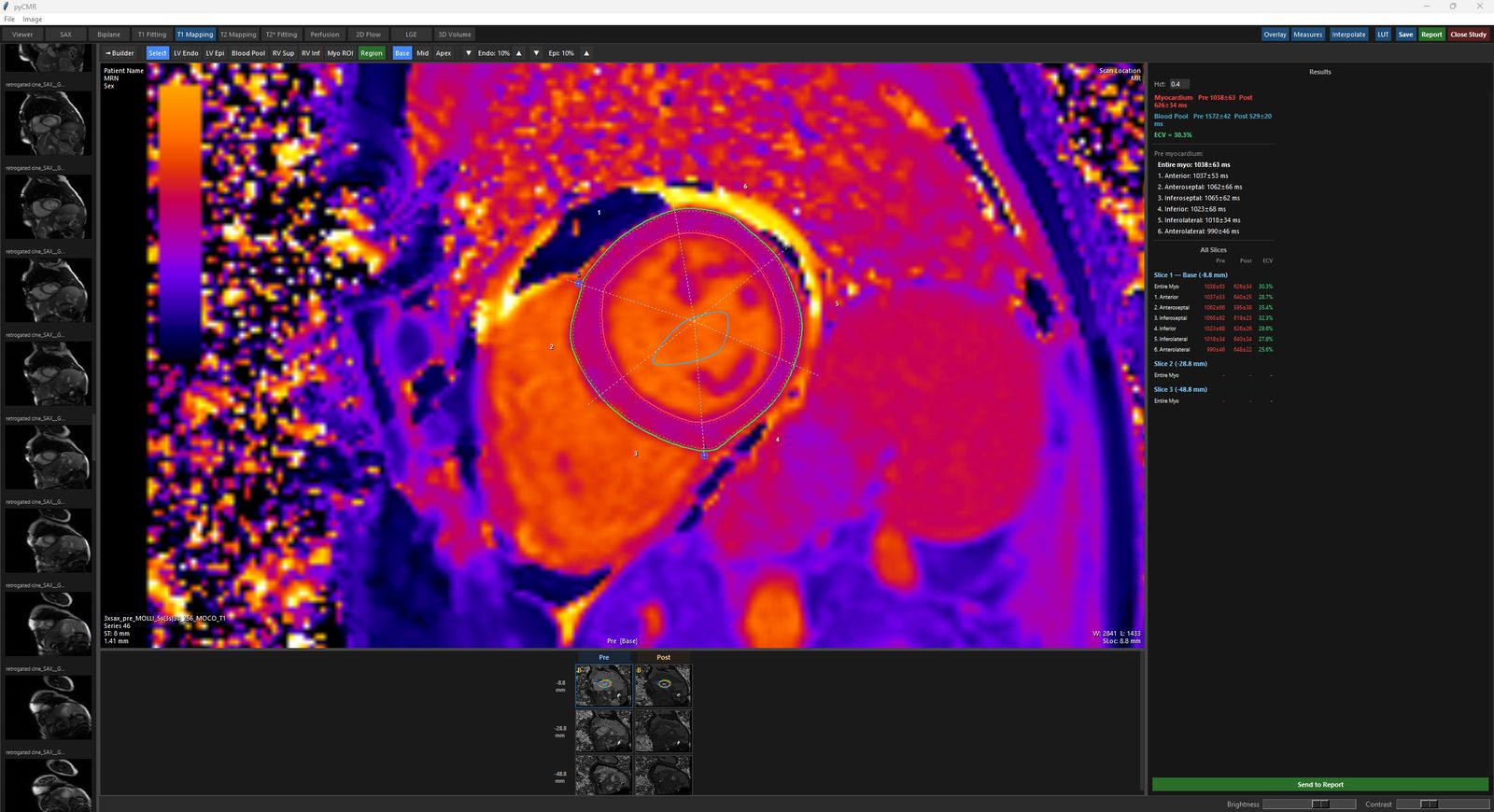
T1 Mapping — from vendor-computed T1 maps ▶
Analysis of a T1 map from the vendor: entire-myocardium and 16-segment AHA T1, with ECV.
- 16-segment AHA T1, tagged Base, Mid, or Apex
- Endo/Epi offset for partial voluming
- Pre/Post ECV per segment
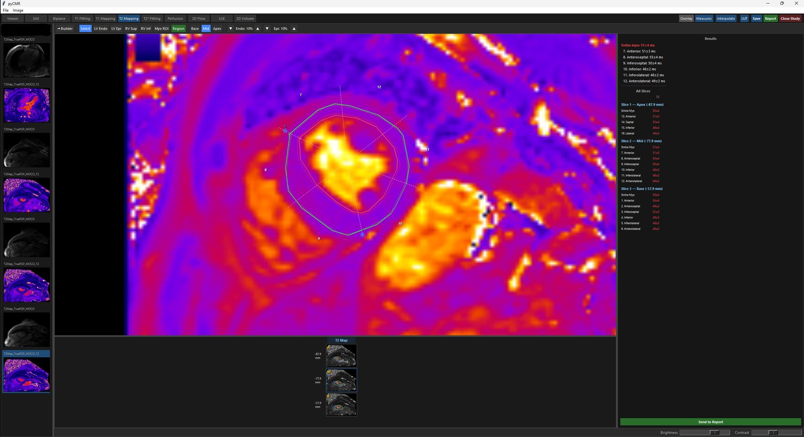
T2 Mapping — from vendor-computed T2 maps ▶
Analysis of a T2 map from the vendor: whole-slice and 16-segment AHA T2 quantification.
- 16-segment AHA T2, tagged Base, Mid, or Apex
- Focal Myo ROI sampling, separate from the segment ring
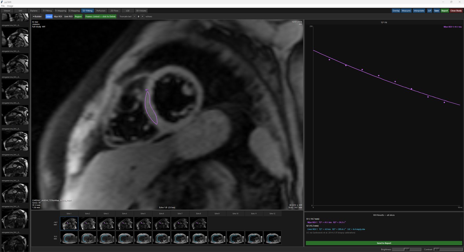
T2* Fitting — cardiac & liver iron loading ▶
Fits a T2* decay curve per ROI from multi-echo source images. Cardiac and liver ROIs.
- Mono-exponential T2*/R2* fitting, with echo truncation
- Cardiac and hepatic iron ROIs
- Liver Iron Concentration via the Garbowski 2014 formula
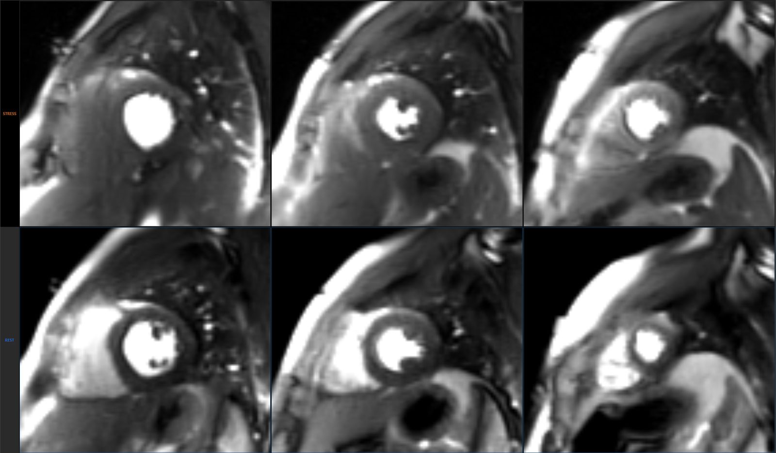
Perfusion — stress & rest perfusion review and defect scoring ▶
Stress and rest first-pass perfusion cine review, side by side, with segment-by-segment defect scoring.
- Independent Stress and Rest playback rows
- 16-segment click-to-grade Stress and Rest bullseyes
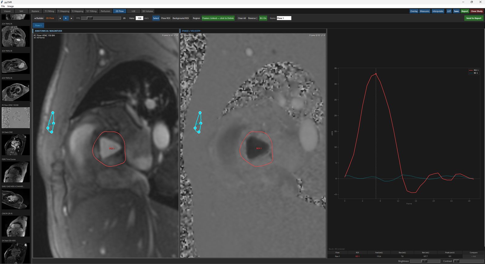
2D Flow — phase-contrast flow quantification and comparison ▶
Velocity-encoded through-plane flow at one imaging level, per cardiac phase, from a Magnitude/Phase series pair.
- Venc auto-detected from vendor DICOM tags
- Background-offset correction from a reference ROI
- Two-ROI comparison for a Qp:Qs shunt ratio
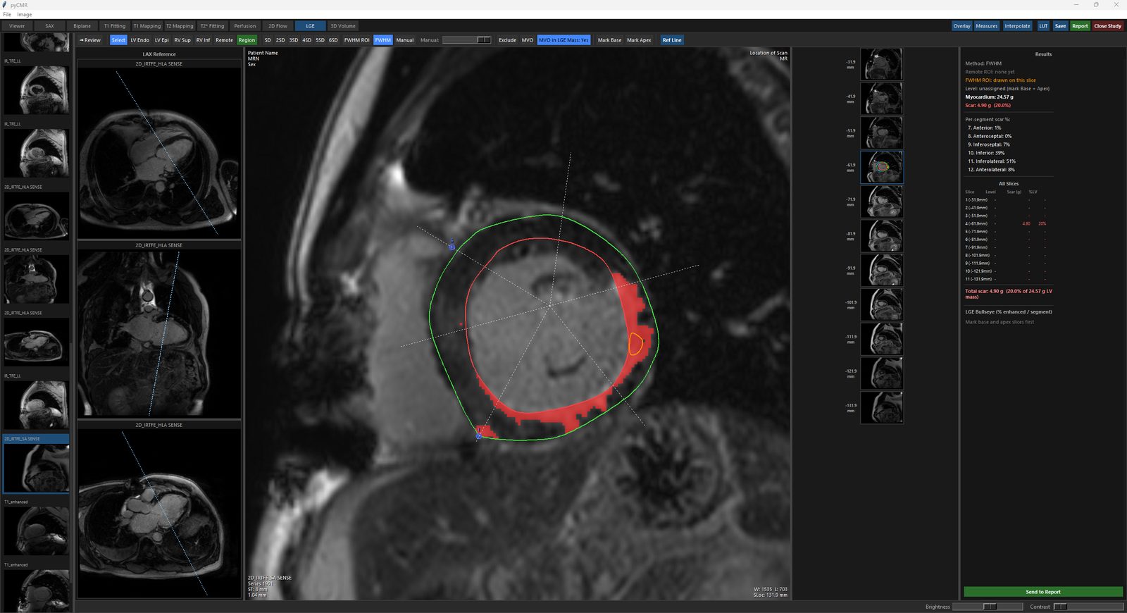
LGE — scar review and quantification ▶
Scar quantification from late gadolinium enhancement imaging, with three interchangeable threshold methods.
- Manual, SD, and FWHM thresholding
- Base-to-apex Remote and FWHM signal carry-down
- Per-segment scar burden and a viability bullseye
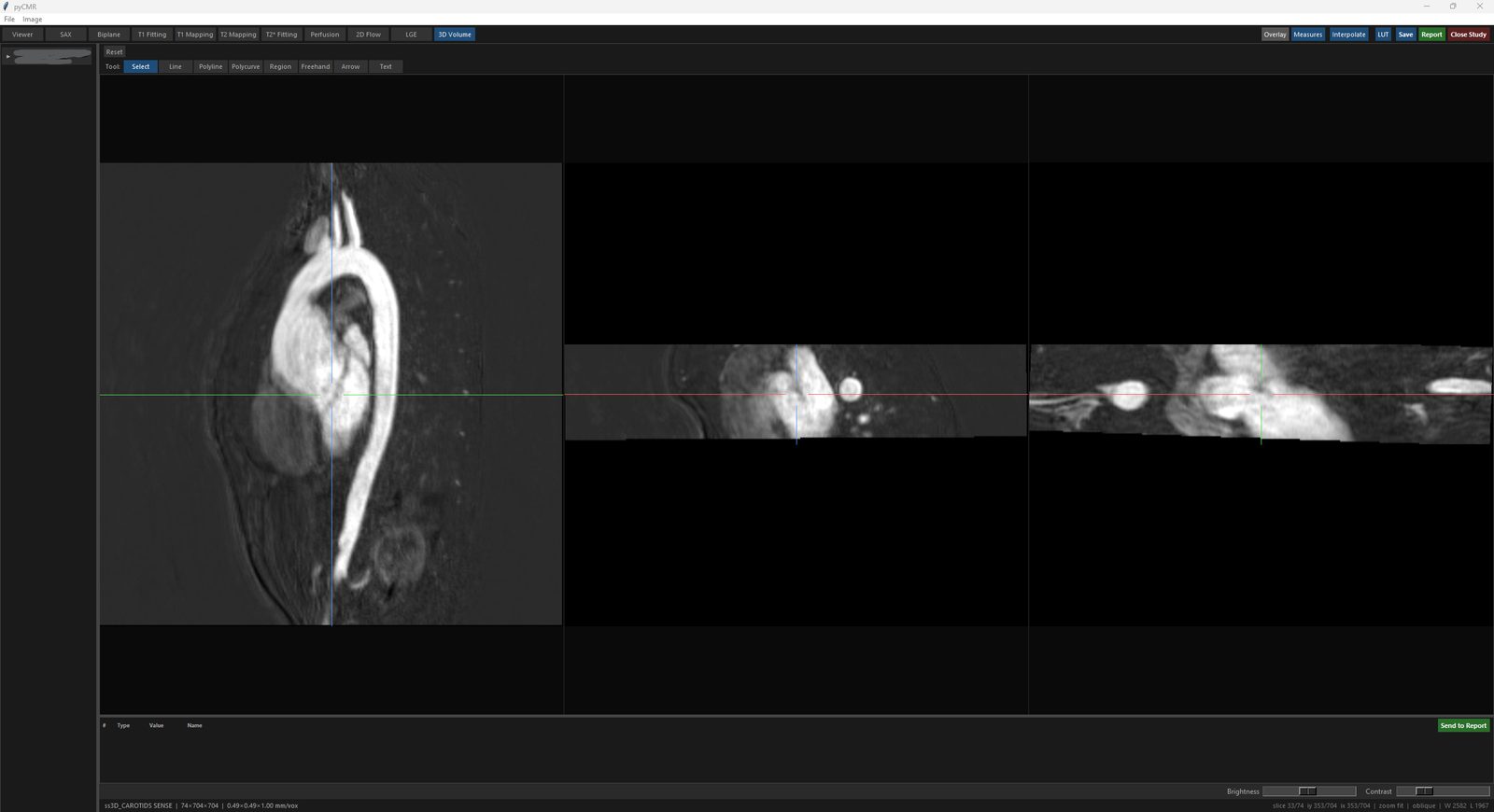
3D Volume — multiplanar reconstruction ▶
Three linked multiplanar (MPR) panels with oblique reformatting, for reviewing 3D whole-heart or angiography.
- Shared crosshair across axial-, coronal-, and sagittal-equivalent panels
- Real-time oblique reformatting by right-click-drag
- Measurements stored as true 3D coordinates